Biophysics of hagfish slime
Hagfishes are bottom dwelling marine animals that are capable of producing startling amounts of defensive slime when they are provoked. We have investigated the composition and mechanical properties of the slime using a variety of techniques. We have also demonstrated that hagfish slime is especially good at clogging the gills of would-be fish predators, which is likely one of its primary functions in life. The slime is composed of fine protein slime threads as well as a mucus component that comes packaged in tiny vesicles. When these components are ejected from the slime glands, they combine synergistically to form a slime mass in which a large volume of water is entrained. Recent work in this area focused on the deployment of both the fibrous and mucus components of the slime in seawater. We now know that the swelling of the mucus vesicles is accelerated by the presence of water pores known as aquaporins in the vesicle membranes. We have also recently shown that unraveling of protein threads from their coiled state involves the dissolution of a seawater soluble glue and the release of strain energy stored in the thread coils. Current research is focused on understanding the mechanisms by which slime threads are manufactured within the cytoplasm of gland thread cells and identifying the proteins involved in slime deployment.
Hagfishes are able to produce vast amounts of defensive slime when they are attacked. Here, a Pacific hagfish in an aquarium is showing off its sliming talents. Photo by Andra Zommers.
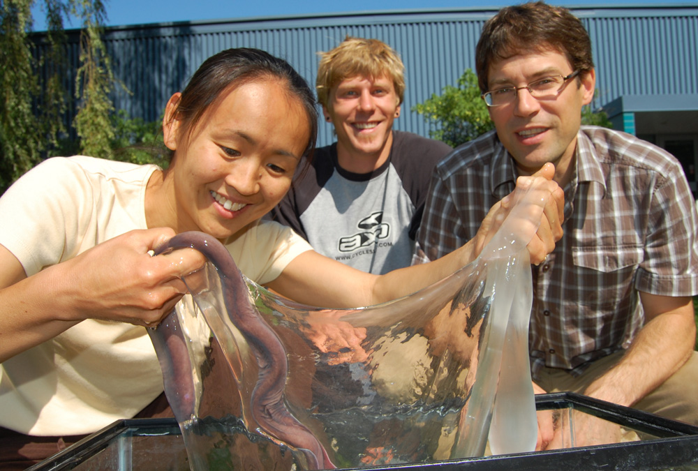
A liter of hagfish slime contains tens of thousands of silk-like protein fibres, each of which is about 15 cm long. Here, bundles of slime fibers can be seen in this slime held up by former M.Sc. student Tim Winegard.
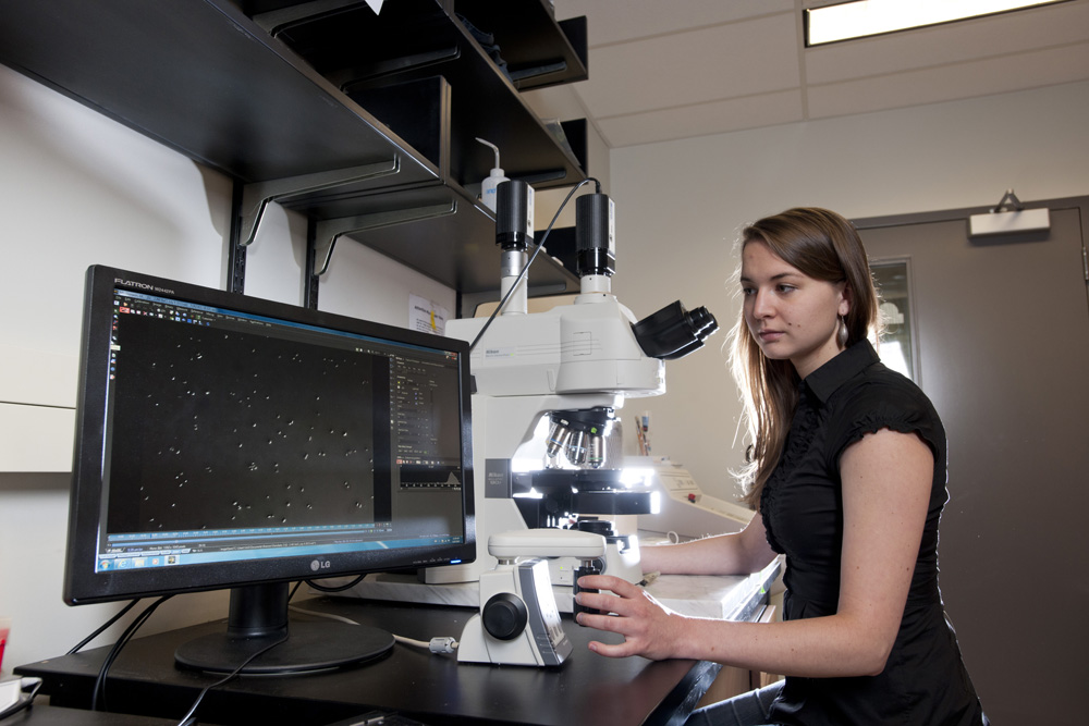
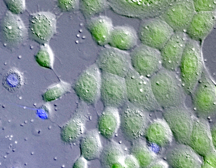
The mucus component of the slime comes packaged in tiny vesicles that are only about 5 micrometers in diameter. Here former M.Sc. student Julia Herr is exploring the behavior of these vesicles under the microscope to understand the transformation they undergo when they are ejected from the slime glands into seawater.
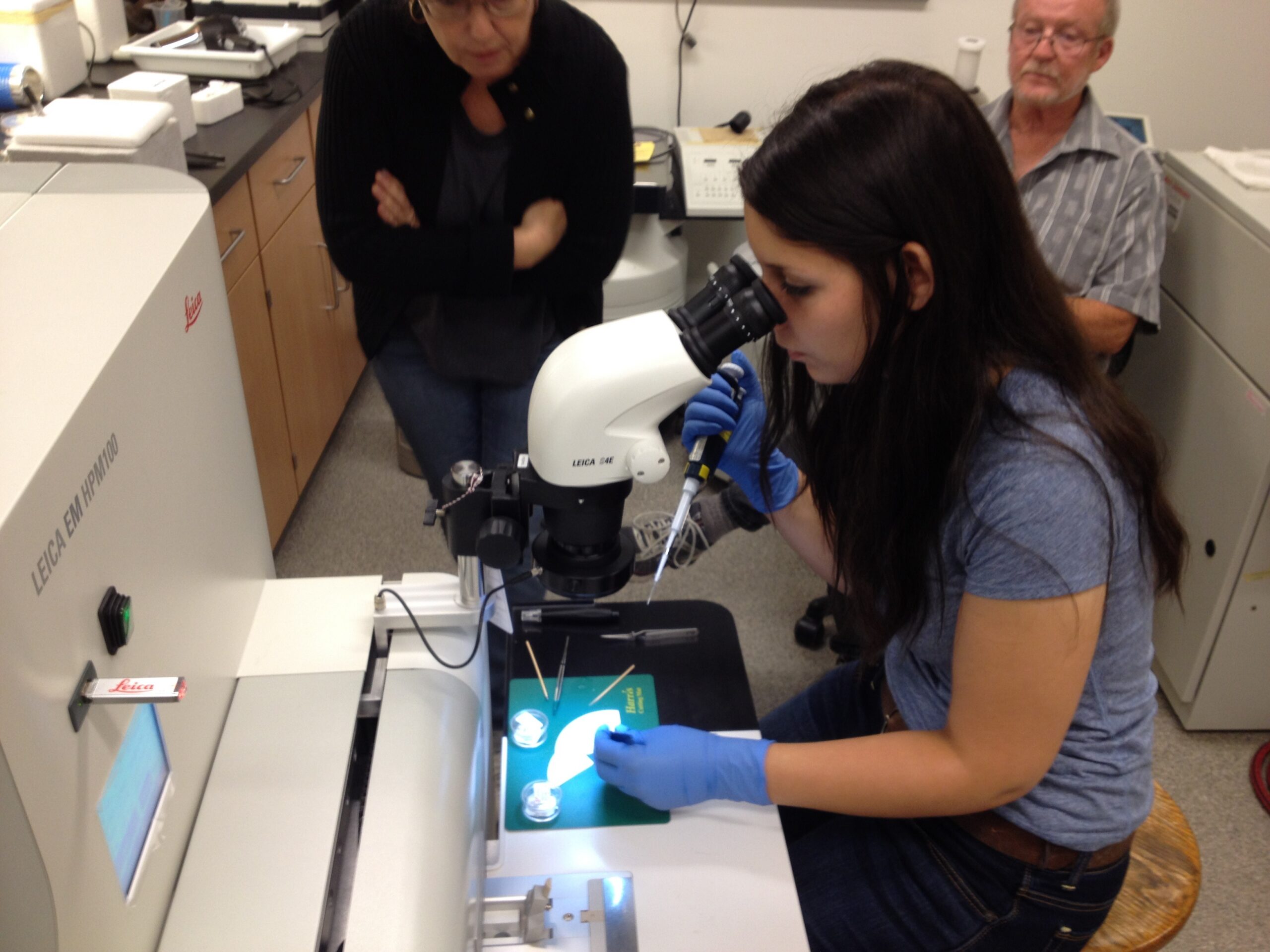
Each mucus vesicle is enveloped by a lipid bilayer that we suspect contains several types of proteins that are involved in the deployment of the vesicles in seawater. Here M.Sc. student Shannon Ferraro is preparing samples for electron microscopy using a high pressure freezing system in the Advanced Analysis Center at the University of Guelph, with help from Bob Harris and Dr. Michaela Strüder-Kypke.
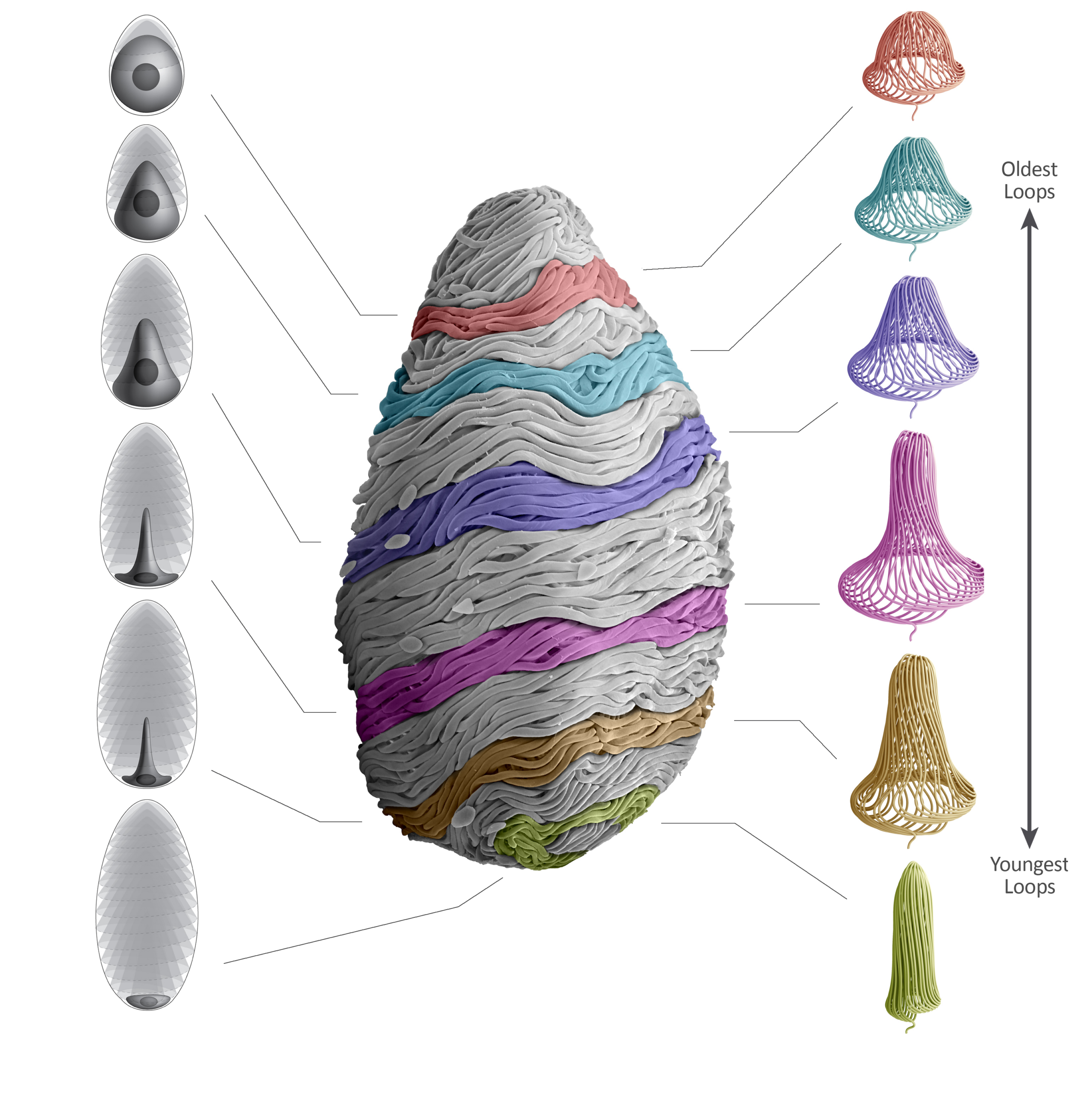
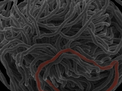
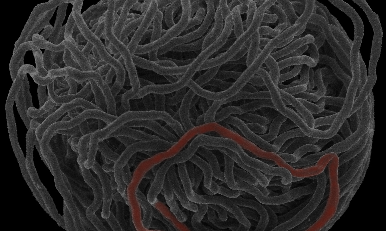
The numerous protein fibers that are found within hagfish slime are produced individually within the cytoplasm of highly specialized cells know as gland thread cells. In a recent paper (Winegard et al. 2014), we elucidated some of the mechanisms involved in the sub-cellular production of these 15 cm long threads.
And here is a movie showing exactly how the slime thread is coiled within a developing thread cell.
And here is a movie of the image stack used to create the 3D model above.
Undergraduates Isdin Oke and Mark Bernards research projects led to a paper that was recently published in the Journal of Experimental Biology. The paper is on the mechanism of unraveling in Pacific hagfish slime thread skeins. You can see a movie of the process below or download the pdf in the Publications section. Mark and Isdin’s work was also featured on the National Geographic website, which you can see here.
Coiled slime threads from Atlantic hagfish do not unravel spontaneously in seawater, and require vigorous mixing to start the unraveling process. Here, a small drop of fresh hagfish slime exudate within a larger drop of seawater is exposed to flow under a microscope to reveal unraveling in some of the coiled threads. Note also how the mucus gets pulled into strands. For more information, see Winegard et al. (2010).
The mucus component of hagfish slime comes packaged in numerous vesicles, which swell and rupture when they are ejected from the slime glands into seawater. Here, former M.Sc. student Julia Herr has captured the rupture of mucus vesicles in vitro under the microscope. If you watch closely, you can see there are two main types of vesicle behaviours – a slow swelling that starts early, and a much faster popping that occurs later in the video. We currently do not understand the significance of having two distinct kinds of vesicles. See both of Julia’s papers for more information (Herr et al. 2010; Herr et al. 2014).
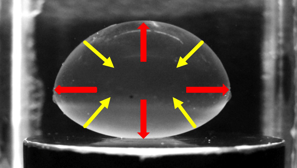
Other Research in the Fudge Lab