Biomechanics of the cytokeratin network in skin cells
Previous research from our lab has demonstrated that the cytoskeletal filaments known as ‘intermediate filaments’ (IFs) are far softer and more extensible than previously believed (Fudge et al. 2003). These findings distinguish IFs from the other two cytoskeletal filaments, F-actin and microtubules, and suggest unique mechanical roles for the IF network in cells. Current research is focused on the role that IFs play in reinforcing epithelial cells against mechanical injury, which has implications for tissue fragility diseases such as epidermolysis bullosa simplex (EBS).
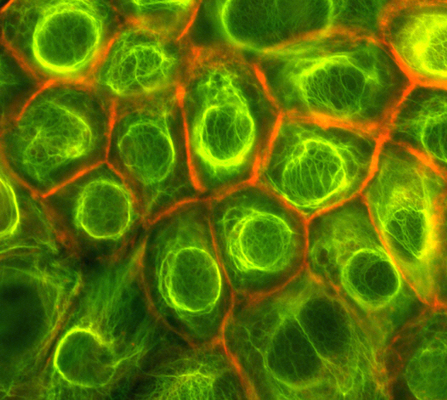
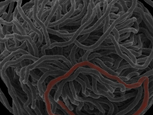
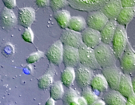
Keratin filaments are the dominant IF type in epithelial cells and they are believed to be critical for the mechanical integrity of epithelia, which is important for their barrier and transport functions. On the left are some human skin cells (keratinoctyes) expressing a green fluorescent version of the keratin protein K14. The cortical belt of actin filaments in these cells has been stained red with the fluorescent actin-specific dye rhodamine-phalloidin.
Our work has demonstrated that the IF network in cells is far more extensible than previously believed and likely acts to protect cells from mechanical injury and returns them to their resting dimensions if they get deformed. For more information, see Kreplak and Fudge (2007), Fudge et al. (2008), and Beriault et al. (2102).
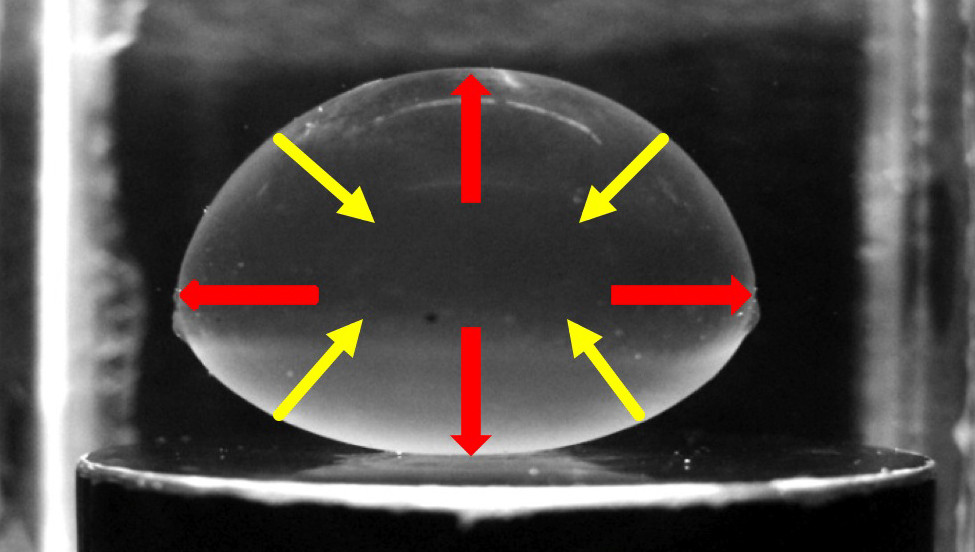
Studying the mechanical behaviour of IF networks in living cells has required us to build a custom cell stretcher incubator that can subject cells to extreme strains while monitoring them under the microscope. To the left is a time lapse movie of immortalized human skin cells grown on a silicone rubber membrane stretched to a strain of 50% and then returned to the resting state.
Other Research in the Fudge Lab