Biomechanics of mammalian keratins
Mammals produce two kinds of keratin materials – soft keratins found primarily in skin, and hard keratins that make up structures such as hair, nail, claw, horn, hoof, and baleen. We are interested in the mechanisms by which mammals produce keratins with such a wide range of morphologies and material properties. One way that keratins differ is in their resistance to hydration, with soft keratins undergoing significant swelling and softening in water, and the hard keratins remaining relatively stiff in water. Within the hard keratins, there is further variability, with rhinoceros horn and echidna quill representing two ends of a spectrum of hydration resistance. We have shown that much of this variability can be explained by the ratio of the two main components of keratins, the keratin filaments and the matrix proteins that glue them together into a functional composite. Keratins like echidna quill have a high ratio of matrix to filaments, with the matrix functioning to resist the swelling and softening of the filaments that would otherwise take place in water. In rhinoceros horn, which has little matrix, the filaments are less restrained and therefore are free to swell and soften in water, making this material quite sensitive to hydration. Our interest in keratin hydration has led us to study whale baleen, which is a fascinating keratin material that must “cure” without the benefit of air drying. We have also shown that mysticete whales boost the stiffness of their baleen via mineralization with calcium nanocrystals such as hydroxyapatite, the primarily mineral in bone.

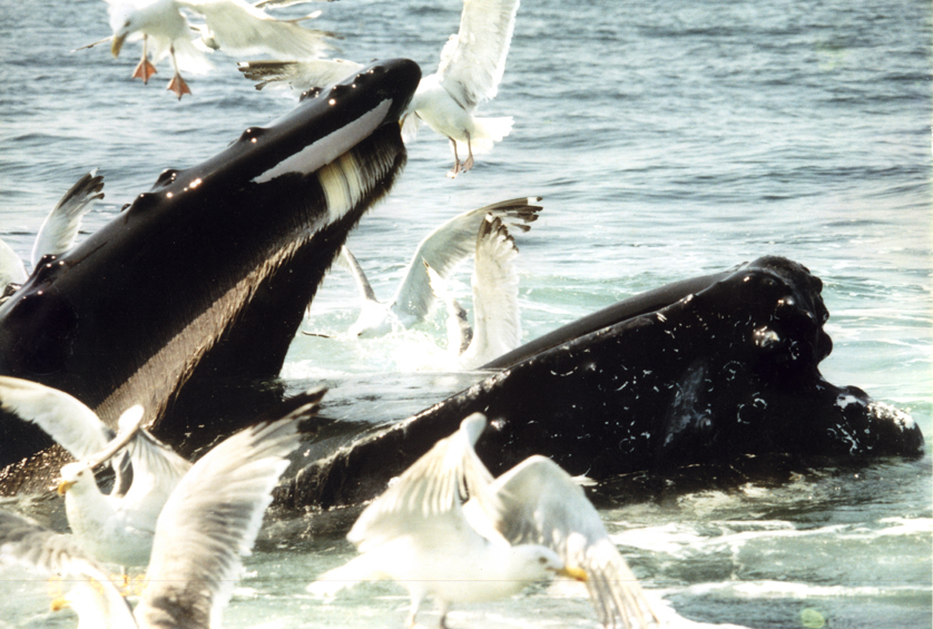
Former M.Sc. student Lawrence Szewciw poses with a few plates of baleen from a humpback whale. Note how the lingual (facing the tongue) side of the plates are frayed into bristles, which form a filtration mat, whereas the labial (facing the lips) side is smooth.

Whale baleen is one of the most heavily calcified keratins, and sei whale baleen is the most heavily calcified of all. Here a micrograph of a sei whale baleen plate prepared with a von Kossa staining protocol reveals the distribution of calcium salts (brown) in and between the tubules that make up the plates and the bristles on the frayed lingual side. For more information, see Szewciw et al. (2010).
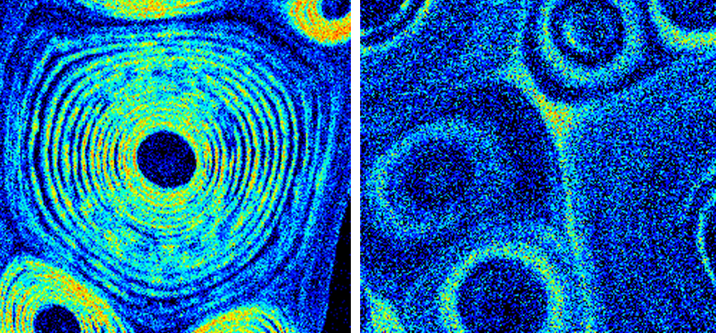
In collaboration with Diane de Kerckhove at the University of Guelph and Geoff Grimes at the University of Surrey, we explored the atomic composition of three species of whale baleen using proton induced x-ray emission (PIXE). On the left is a heat map showing the distribution of calcium salts (with warmer colours representing higher concentrations) in sei whale baleen, and on the right is minke baleen. For more information, see Szewciw et al. (2010).
Other Research in the Fudge Lab