Biophysics of the ocular lens
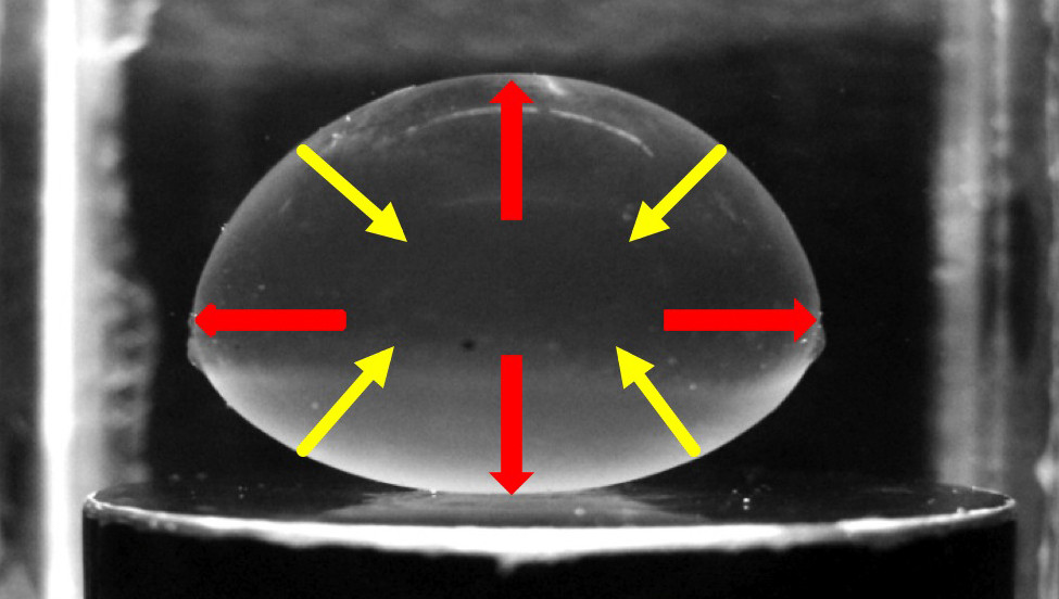
The vertebrate ocular lens consists mainly of long and skinny cells known as fiber cells. These unique cells contain cytoskeletal filaments known as “beaded filaments,” which are heteropolymers of the proteins filensin and phakosin. In humans, mutations in beaded filament genes have been linked to autosomal-dominant congenital cataracts (ADCC). Because phakosin and filensin both belong to the intermediate filament gene family, we suspected that beaded filaments play a mechanical role in the lens. With collaborators John Hess and Paul FitzGerald at UC Davis, we showed that mouse lenses lacking beaded filaments are softer than lenses from wild type mice, suggesting a mechanical role for beaded filaments in the lens. In a recent collaboration with G-J Won and Vivian Choh at the University of Waterloo, we showed that the actomyosin network within chick lens cells also plays a role in setting the mechanical properties of the lens.
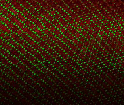
In a recent paper in Molecular Vision, GJ Won demonstrated that disruption of actomyosin networks in lens fiber cells causes a softening of the chick lens. In this confocal image, the highly organized actomyosin lattice can be seen in these hexagonal cells (green, F-actin; red, myosin).